Diagnostic Testing & Imaging
An EKG, or electrocardiogram, is a simple, painless test that measures and records the heart�s activity. It allows us to monitor the heart�s health and detect other heart issues. Tiny patches called electrodes are strategically placed on the patient�s chest, arms, and legs. The electrodes are connected to the EKG machine by lead wires, and detect the heart�s electrical signals. The results show on the monitor as a moving line, and are printed for our doctor to review and interpret.
Make an AppoinmentAnkle-brachial Index (ABI)
Ankle-brachial index?(ABI) is a test for PAD, that compares the blood pressure in the leg to the blood pressure in the arm. If the blood pressure in the arm is higher than the blood pressure in your legs, this could be an indication of decreased blood flow in your legs known as Peripheral Artery disease or PAD.
EKG (Electrocardiogram)
An EKG, or electrocardiogram, is a simple, painless test that measures and records the heart�s activity. It allows us to monitor the heart�s health and detect other heart issues. Tiny patches called electrodes are strategically placed on the patient�s chest, arms, and legs. The electrodes are connected to the EKG machine by lead wires, and detect the heart�s electrical signals. The results show on the monitor as a moving line, and are printed for our doctor to review and interpret.

Echocardiogram
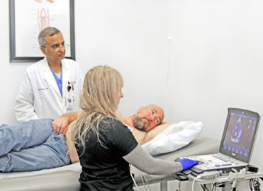
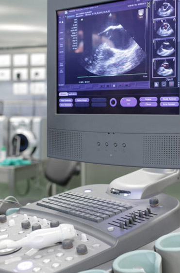
An echocardiogram uses high frequency sound waves to produce pictures of the heart. The ultrasound technician guides a small hand-held device called a transducer over the chest, that emits soundwaves. These sound waves bounce off internal substances and structures (in this case the heart), creating echoes that are picked up by the transducer. These are converted into moving images of the heart�s chambers, valves, blood vessels and pumping action. Combined with doppler technology, the speed and direction of the heart�s blood flow can also be measured at the same time.
Echocardiograms can help detect many aspects of heart disease including tumors, abnormal holes between the heart chambers (ASD & PFO), blood clots, and issues with the heart�s blood vessels, valves and pericardium (the heart�s outer lining).
Stress Echocardiogram
A stress echocardiogram, or stress echo test, assesses how well the heart is functioning when it is working harder due to physical stress. For comparison reasons, a resting echocardiogram is first administered. An ultrasound technician guides a small hand-held device called a traducer over the chest. It transmits soundwaves that are converted to moving images of the heart on the computer screen. Next, the patient walks on a treadmill while the heart rhythm and blood pressure are monitored. When the heart rate increases to a certain point, a second set of echocardiogram images are taken. This test shows how efficiently your heart pumps blood, can detect heart issues that may not show up at rest, and can measure the severity of heart issues such as a blockage. In cases where the patient is unable to walk on a treadmill, an IV medication can be given instead that will mimic the effects of exercise to increase the heart rate.
Echo Bubble Study
An echo bubble study can be performed during an echocardiogram. During the echocardiogram, a saline solution is rapidly mixed with air to produce tiny bubbles, and is then injected into the patient�s vein in their arm through an IV. The bubbles circulate up the vein into the right side of the heart, showing up on the echocardiogram. The bubbles should normally then be filtered out by the lungs. However, if these bubbles appear on the left side of the heart, this indicates that there is a hole (an ASD or PFO) between the heart�s two upper chambers (the left and right atria).
Holter Monitor
A Holter monitor is a portable device worn by that patient that records the heart beat and monitors for irregular heart rhythms (arrhythmias). The Holter Monitor is attached to the chest with round patches called electrodes. It records the heartbeat continuously for 24 to 48 hours as the patient goes about their day.
Event Monitor
An Event Monitor is a portable device worn by the patient that records the heart beat and monitors for irregular heart rhythms (arrythmias.) The Event Monitor is attached to the chest with round patches called electrodes. It is similar to the Holter monitor, except that the event monitor only records the heartbeat when activated. Some event monitors will start recording when an abnormal heart rhythm is detected. It also records much longer, for a month or more.
Certain irregular heart rhythms can occur infrequently, and therefore may not be picked up by a standard EKG performed at our clinic. The Holter Monitor and Event Monitors are better options in this case since they record over longer periods of time.
MUGA Scan
A multigated acquisition (MUGA) scan is a nuclear imaging test that creates video images of the lower chambers of the heart, called ventricles, to check how well they are pumping blood. The MUGA Scan measures the patient�s ejection fraction (EF). The ejection fraction is the percentage of blood that is being pumped out of the heart each time it beats. The standard percentage is between 50 to 75%. During the test, the patient is attached to an EKG machine to measure the heart rate. Then, through an IV, the patient receives an injection of a radioactive tracer that attaches to the red blood cells. This allows the camera to take pictures of the blood in the ventricles, which are responsible for the pumping of blood. It shows any abnormalities in the size of the ventricles and in the movement of blood through the heart. This test is often done first when you are resting, and then again after walking on a treadmill. This will show how well your heart is able to pump when working harder. Other names for this test include cardiac blood pooling imaging, nuclear heart scan, nuclear ventriculography, and radionuclide ventriculography.
Abdominal Aortic Aneurysm Screening
What is an Abdominal Aortic Aneurysm?
The aorta is the main artery in the body that brings oxygenated blood from the heart to all organs. An abdominal aortic aneurism (AAA) is a condition when an area of the lower part of the aorta becomes enlarged. If the aorta ruptures, this can cause life-threatening internal bleeding. An AAA is often asymptomatic and can gradually grow for a long period of time undetected. Males over 65 years old, current or previous smokers, and those with a family history of AAA have an increased risk.
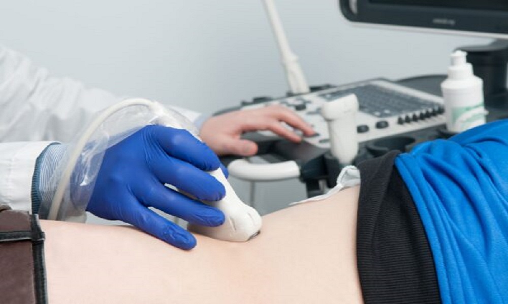
Abdominal Aortic Ultrasound
An abdominal ultrasound is a painless diagnostic imaging test performed that is commonly used to diagnose an abdominal aortic aneurism. During the test, the ultrasound technician guides a small hand-held device called a transducer over the abdomen where the aorta is located. The transducer emits high frequency sound waves that echo off the organs and internal structures in the abdomen, including the aorta, and convert into images on a computer screen. Combined with Doppler technology, the speed and direction of the blood flow through the abdominal organs and structures can also be assessed.
Patient Instructions: Abdominal Aortic Ultrasound

Arterial Duplex Ultrasound / Arterial Doppler Ultrasound
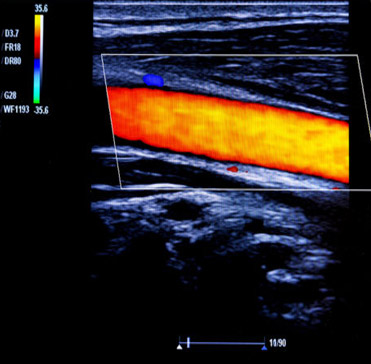
An arterial duplex ultrasound, also known as an arterial doppler ultrasound, shows blood flow through the arteries. The ultrasound technician guides a small hand-held device called a transducer over the area being examined. The transducer emits high frequency sound waves that echo off moving objects, such as red blood cells, and convert into images on a computer screen. This allows us to measure blood flow in the arteries to see if there is a blockage. Based on the findings of this test, your doctor will determine if further testing or treatment are necessary.
Venous Duplex Ultrasound / Venous Doppler Ultrasound
A venous duplex ultrasound, also known as a venous doppler ultrasound, shows blood flow through the veins. The ultrasound technician guides a small hand-held device called a transducer over the area being examined. The transducer emits high frequency sound waves that echo off moving objects, such as red blood cells, and convert into images on a computer screen. This allows us to measure blood flow in the veins to see if there is a blockage. Based on the findings of this test, your doctor will determine if further testing or treatment are necessary.
Carotid Ultrasound
A carotid ultrasound is a painless diagnostic imaging test that examines the carotid arteries, which supply blood to the brain, face and neck. It checks for blood clots and plaque buildup in the carotid arteries, which could lead to a potential stroke. The technician guides a small hand-held device called a transducer over each side of the neck where the two carotid arteries are located. The transducer produces high frequency sound waves that echo off of the structures in the neck and convert into images on a computer screen. Combined with doppler technology, the speed and direction of the arterial blood flow can also be measured at the same time.
Renal Artery Ultrasound
A renal artery ultrasound is a painless diagnostic imaging test that examines the renal arteries, which supply oxygenated blood from the heart to the kidneys. It checks for renal artery stenosis, the narrowing or blockage of the arteries due to a buildup of plaque. This can lead to high blood pressure and decrease in kidney function. The kidneys require sufficient blood flow to filter waste and eliminate excess fluid. During the test, the ultrasound technician guides a small hand-held device called a transducer over the abdomen where the renal arteries are located. The transducer emits high frequency sound waves that echo off the renal arteries and convert into images on a computer screen. Combined with Doppler technology, the speed and direction of the arterial blood flow can be assessed at the same time.